
Breast Imaging & Diagnostic Services
Learn about our comprehensive breast imaging options and what to expect during a mammogram.
Latest Technology For The Most Accurate Results
One in 8 women will receive a breast cancer diagnosis in their lifetime, making early, accurate detection vital. At Stamford Health's Breast Center, we offer the most advanced diagnostic and imaging technology, including mammography, to detect any abnormalities in the breasts.
A health care provider (an OB-GYN or primary care provider) will help determine which test is right for you based on your unique breast health needs. If you are diagnosed with breast cancer, Stamford Health will be here for you throughout every step of the journey. Through our collaboration with Dana-Farber Brigham Cancer Center, you’ll have access to the latest clinical trials, virtual second opinions, and more. Learn more about our comprehensive services below.
What To Know About 3D Mammography
Stamford Health is proud to have been the first breast center in the region to offer 3D mammograms to everyone.
A 3D mammogram, also known as tomosynthesis, is faster and uses less radiation than a standard film mammogram. Studies have shown that 3D mammography is better at detecting invasive cancer while limiting unnecessary biopsies and false positives. More definitive images and greater accuracy mean less time and worry for you.
-
When and how often should you get a mammogram?
Mammography detects breast cancer long before it can be seen or felt in your breast. The current screening recommendations for people of average risk (meaning they do not have risk factors that equate to an increased risk for breast cancer) include:
- Age 20-39: Regular breast self-exam or clinical breast exam every two to three years.
- Age 40: Schedule a routine baseline mammography screening and then repeat each year.
- Elderly women: Continue annual screening for as long as you are reasonably healthy and would be a candidate for breast cancer treatment.
-
What is a breast MRI? Why might you need one?
Compared to mammography and ultrasound, breast magnetic resonance imaging (MRI) is a completely different way of looking at the breast. It is an advanced tool using MRI technology, sophisticated computers, and 3D techniques that look deeply into the breast to discover abnormalities that might not be visible in other exams.
The following groups may require an annual breast MRI screening:
- Women who have a BRCA gene mutation or have a first-degree relative (mother, sister, or daughter) with a BRCA mutation.
- Women who have received radiation treatment to the chest between the ages of 10 and 30.
- Women who have a rare medical condition linked to breast cancer (Li-Fraumeni, Cowdon, or Bannayan-Riley-Ruvalcaba syndromes) or have a first-degree relative with one of these syndromes.
- Women with a 20-25% greater lifetime risk of developing breast cancer.
- Other reasons for getting a breast MRI include current breast cancer diagnosis, prior to breast cancer surgery, and to detect a breast implant leak.
Breast Imaging Services
Stamford Health’s Breast Center offers the most cutting-edge technology that can be performed quicker and with greater accuracy.
Think You Might Be At An Increased Risk For Breast Cancer? We Can Help.

Our team of breast specialists will determine your risk after a thorough family and medical history intake and a breast exam. If you need additional care, our breast navigators will come up with a care plan created specifically for you.

Our Nurse Navigator Program

-
How Do you Prepare for a Breast MRI or mammogram?
For a breast MRI, normal daily routine is fine. Please do not wear any jewelry.
For a mammogram, do not wear deodorant, talcum powder, or lotion under your arms or on your breasts on the day of your exam as they can appear as calcium spots on your mammogram.
If you have had previous mammograms at another location, please be sure to bring your past images with you so that the radiologist can compare them successfully. We’re also happy to obtain any past images for you.
-
How Long Will It Take for you to See the Results of your Exam?
When your exam result is available for viewing will vary depending on the type of imaging. Patients with normal mammogram results will be notified via MyChart. This means you’ll get your results faster — right after your images are reviewed. If you don’t have a MyChart account, your results will be mailed to you. If you have abnormal results, you’ll be contacted by phone about next steps.
-
What is the difference between a screening and diagnostic mammogram?
A screening mammogram is performed to detect breast cancer in women who have no apparent symptoms.
A diagnostic mammogram is used to more closely review the breast tissue, typically following symptoms or after a screening mammogram showed abnormal or suspicious results.
-
What Should You Expect When Getting a Breast MRI?
Prior to the exam, an IV will be inserted into your arm to administer the contrast material. You will lie face down and very still with your breasts resting in a cushioned coil while you are moved in and out of the MRI machine. Halfway through the exam, you will receive a painless injection of contrast and more images will be taken.
If you are claustrophobic or anxious about your MRI exam, you may want to contact your physician at least two days prior to your appointment to obtain a prescription for a mild sedative.
After the exam is complete, the radiologist will read and interpret your MRI and your physician will be contacted with the results.
-
Do You Still Need to Have a Yearly Mammogram if you Get a Breast MRI?Absolutely. A breast MRI should not take the place of your annual mammogram. If you are at high-risk, you should have an annual breast MRI in addition to your annual mammogram.
-
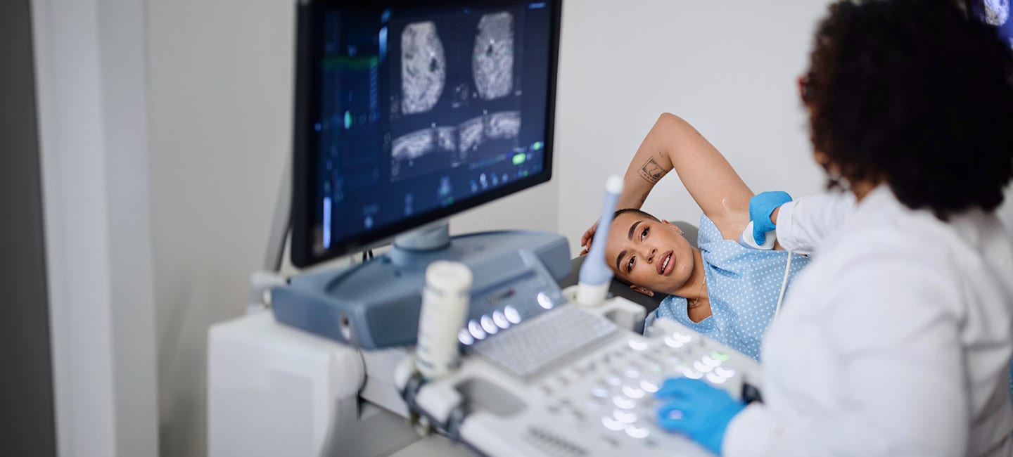
If you have dense breasts and need both mammography and breast ultrasound, are they covered by insurance?
For most patients with private insurance, screening breast ultrasounds will be covered by insurance.
As of October 2023, Medicare will no longer cover breast ultrasounds used for screening patients with dense breast tissue, if the sole indication for the exam is dense breast tissue. Due to this change, Medicare may not cover your breast ultrasound.
While mammography is a powerful screening tool in the early detection of breast cancer, it is less effective in patients with dense breasts. Research has found women who have dense breast tissue (which can hide small abnormalities in the breast) benefit from supplementary screening tests which includes breast ultrasound. Therefore, we continue to recommend screening breast ultrasound in those with dense breast tissue to increase sensitivity in detecting breast cancer.
If a patient wishes to proceed with the ultrasound, understanding it may not be covered, they will need to sign an advanced beneficiary notice (ABN) before the ultrasound is done. The rates for screening breast ultrasounds range between $115 to $215, dependent on the study being performed.
Patients can reach out to our Contact Center with any questions: 203.276.2602.
Pregnancy And A Breast Cancer Diagnosis
When Alexandra Belluzzi found out she was pregnant after two miscarriages, she was filled with excitement and hope. However, when a rash appeared on her left breast and only worsened with time, her obstetrician ordered a breast ultrasound, which led to biopsies. Her doctor then delivered a devastating diagnosis: breast cancer. “I didn’t move. I didn’t say anything. I didn't know what to do,” Belluzzi said. “It felt like good things were coming, and this just totally knocks you off your feet.”
Under the care and guidance from her oncologist, Dr. Paul Weinstein, who was in constant communication with her obstetrician, Belluzzi began chemotherapy before delivering her baby early via C-section. Throughout her treatments, though, she always kept her thoughts on what matters most. “I wanted to be there for my daughter,” she says. “That was it.” Find out how the rest of her journey unfolded in the video here.